AI-assisted High-Framerate Speckle Image Analysis
PhD - Gent | More than two weeks ago
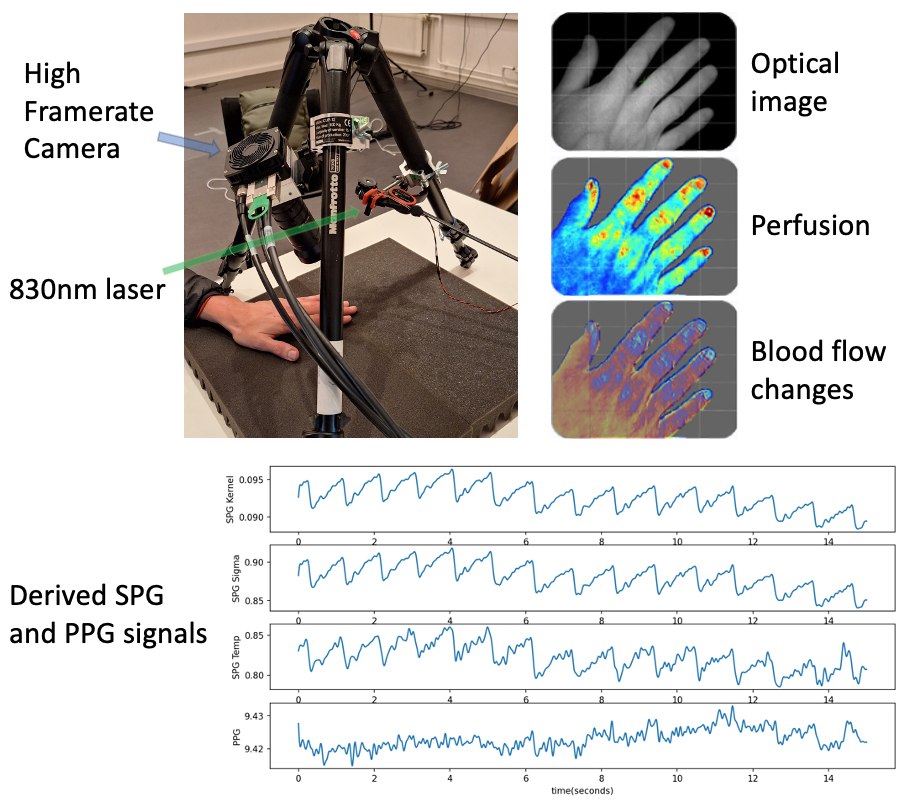
Photoplethysmography (PPG) is an optical method that measures blood volume variations over time by detecting changes in non-coherent light absorption. It provides valuable information on heart rate, peripheral oxygen saturation (SpO2), and can serve as a proxy for blood pressure. Even though the above-mentioned technique has been extensively described in the literature, the actual clinical implementation is hampered due to low signal quality in people with skin melanin, low perfusion, or during varying ambient light conditions. Speckle plethysmography (SPG) presents a promising alternative, this method is based on Laser Speckle Contrast Imaging (LSCI), utilizing speckle patterns that are created on the detector when coherent light interacts with a tissue. Both SPG and LSCI quantify speckle blur due to movement of scatter particles (e.g. red blood cells). Unlike PPG, SPG is inherently less affected by changes in ambient light and skin melanin, making it a more reliable option for diverse clinical settings.
IMEC has recently introduced a novel concept of High-Framerate Speckle Imaging, where laser speckle patterns are recorded with high-framerate camera, taking 3000 framed per second (fps) in full HD resolution, as opposed to 30 fps in conventional LSCI. High-framerate imaging allows us to capture vital signals (such as parts of cardiac cycles, local tissue perfusion and oxygenation changes, blood velocity, etc.) with high signal quality above current state of the art (PPG). In addition, very fast changes and functions of tissue to explore extraction of additional information: a) separation if vessel wall motion from blood flow velocity, b) analysis of viscoelastic behavior and c) to quantify absorption and scattering properties of tissue. High image resolution allows us to simultaneously compare multiple tissue types and to localize the analysis on each tissue type separately. Laparoscopic surgery is one of the cases that could benefit from HFSI. The success of laparoscopic surgical procedures depends to a great extent on good tissue oxygenation (anastomotic leakage, which occurs in approximately 1 of 6 patients and carries up to 28% mortality rate) and the existing techniques are not able to assess whole tissue oxygenation and function during surgery. Perfusion and functional imaging prove critical in surgeries involving blood vessel manipulation such as Coronary Artery Bypass Grafting (CABG) and oxygenation measurements can accurately detect death heart tissue, which is all achievable in a non-invasive way using HFSI, unlike the current state-of-the-art methods which are invasive and cumbersome.
The high temporal and spatial resolutions of HFSI bring challenges as well. Due to low integration times, the images have low signal-to-noise ratio (SNR) and full HD resolution combined with 3000 fps imaging creates large amounts of data in short periods of acquisition (approximately 1 GB per second), which brings forth the necessity of efficient data processing. Clinical applications will require near-to-real time acquisition and data processing (in order to provide the feedback to the physicians and help guide the surgical procedure and prognose the outcome), while combined with accurate local data analysis to allow for distinction between various tissue types (e.g. soft tissue, blood vessels, lesions).
The Photonics-for-health Group (part of AI & A) in IMEC-NL has vast experience in development of speckle imaging for a variety of application fields like biomedicine, automotive consumer electronic. The Image Processing and Interpretation group of IMEC has been working on a number of biomedical and medical image processing and analysis applications and has a long-standing experience in the field. The Experimental Surgery Research group of Ghent University Hospital (the clinical partner) focuses on small and large animal models of colorectal and ovarian cancer and has considerable experience with the design, GCP compliant execution, and analysis of investigator-driven clinical trials with translational research endpoints. Soft Tissue Surgery group (Small Animal Department) of Faculty of Veterinary Medicine at Ghent University conducts clinical scientific research related to small animals of multidisciplinary character. International collaboration on this topic includes the Institute of Cardiovascular Diseases in Sremska Kamenica (Serbia), focusing on research in cardiovascular surgery.
We are looking for a motivated Ph.D. Candidate who is interested in pushing the HFSI technology to clinical practice. You will: (1) conduct experiments to design the optimal HFSI setup for various surgical procedures (open-body and laparoscopic in-vivo imaging) (2) develop novel image processing and analysis methods using AI-based tissue modelling, segmentation and classification to allow for efficient tumor detection, critical structure identification, tissue oxygenation and perfusion measurements and (3) validate the whole setup on phantoms, animals and patient studies.
Required background: Computer Science or equivalent
Type of work: 70% modeling/simulation, 20% experimental, 10% literature
Supervisor: Hiep Luong
Co-supervisor: Evelien Hermeling
Daily advisor: Danilo Babin
The reference code for this position is 2026-147. Mention this reference code on your application form.